Wednesday, August 12, 2009

Ant Night

We presented the Nano Gigapan ant images at the California Academy of Sciences Nightlife. Nightlife is a evening social event at CAS that happens every Thursday evening. It gives people a chance to explore the museum at night and ask questions. Nano Gigapan set up a table and had the opportunity to present some of our images and talk to people about our work.

Aphaenogaster occidentalis

This Nano Gigapan is of an ant commonly found in Northern California. We found this one in the kitchen. It is the same species as the one below that's holding a fly.

This Nano Gigapan is a head shot of the ant that is composed of 80 pictures. The ant is magnified 500x.

You can see optical images of it on ant web. http://www.antweb.org/description.do?rank=species&name=occidentalis&genus=aphaenogaster&project=calants

View the full image at GigaPan.org

Monday, July 20, 2009

The Nano GigaPan in Action

In this video you can see how the Nano Gigapan is hooked to the SEM, how it moves the stage, as well as how the pictures are taken. Jay was explaining the process while taking a Nano Gigapan himself. If you want to see the finished image that he was taking during this video.

Thursday, July 16, 2009

Ant holding a Fly

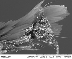
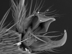
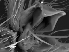
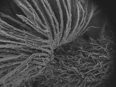
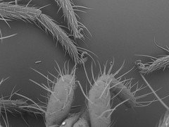
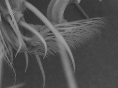
This is an SEM Nano Gigapan of an ant holding a fly in its mandible. This image came about when we found some ants this morning in the kitchen and decided to take them into work to image. While looking for other cool things to image I also stumbled across a very small fly that was dead on the table (you might not believe me, but the house we're staying at is actually really nice, and at least appears very clean). Lacking another container for putting samples into, we dropped the fly in with the live, albeit confused ants, saying "they probably wont eat it." Seconds after touching the bottom of the container, the ant you see here snatched the fly up and proceeded to hold on to it for not only the commute into the office, but also during a stint in the freezer, a move from the container to the SEM stage, and then while in a vacuum. That is dedication.

The ant and fly are magnified 400x and this image is composed of 288 pictures taken with the SEM.


View the full image at GigaPan.org

Wednesday, July 15, 2009

Match Heads


This Nano Gigapan shows two match heads side by side, illustrating the difference (as seen under a scanning electron microscope) between a unlit strike anywhere match and one that has been burned. This difference is clear with the naked eye, but can you tell which has been burned looking at it with a different tool? This image is composed of 140 pictures of matches magnified 150x.

View the full image at GigaPan.org

Tuesday, July 14, 2009

Drift wood & Pygmy forest twig

Here are two gigapans of two different wood samples. Neither of these are quite the bark that someone requested, however, they both look pretty nifty. This first nanogigapan is of a piece of drift wood, the image is made up of 25 pictures taken using a scanning electron microscope. This piece of driftwood was found on the Northern Californian coast. The segment we imaged is of the very center of the wood and you can see this by looking at how the structures seems to radiate from the middle of the gigapan. The driftwood is magnified 300x.

View the full image at GigaPan.org

The second nanogigapan is of a cross section of a twig taken from a pygmy tree in Mendocino. The twig is magnified 250x and you can make out the growth rings in this image.

Rollie Pollie

This is the latest nanogigapan and is an image of a pill bug, or rollie pollie. This view is of the rollie pollie unrolled and on its back (a position that was rather hard to prepare because when faced with premature death the rollie pollie rolls up in to a small ball). Because the specimen was so large, I was only willing to magnify it 200x so that I only had to take 250 images. However, even with relatively small magnification it is a fun image to explore, especially because it is of such a familiar insect.


View the full image at GigaPan.org

Friday, July 10, 2009

NanoGigaPan project works with STAR Participants


The project is currently working with Lisa Adams, a Student Teacher and Researcher (STAR) participant, who is spending the summer at NASA Ames. Adams intends to design five different lesson plans that use Nano Gigapan images to help students more easily grasp and visualize the material. So far we’ve imaged pollen, a pill bug, and a cockroach leg by her request. Hopefully this will help expose school aged children to the world of the small and show them how much is out there that they can’t even see.

Wednesday, July 8, 2009

Barnacle Gigapan

This barnacle Nano Gigapan is really cool. Take your time, really zoom in and explore this one. The barnacle was found washed up on the back of a crab shell at Mendocino's big river beach. In this Nano Gigapan you can see the crab shell around the base of the barnacle.

This image is composed of 384 pictures taken with a scanning electron microscope, which took me around 5-6 hours to capture. The barnacle is magnified 800x.


View the full image at GigaPan.org

Tuesday, June 30, 2009

The Latest Nano GigaPant

This ant is from Madagascar, and is named Eutetramorium mocquerysi. The species is notable for having wingless queens that are indistinguishable from workers.

This image is composed of 400 pictures magnified 400x using a scanning electron microscope. The ant was given to us to image by Brian Fisher an entomologist at the California Academy of Sciences.

For some more information, along with optical microscopic images click here.


View the full image at GigaPan.org

Wednesday, June 24, 2009

More Ant Nano Gigapans!

More Ants are on their way! The Nano Gigapan project has received some ant samples from colonies at the California Academy of Sciences which are in the process of being imaged. Seeing as there are over 30,000 species of ants in the world, we could have a new ant image every day for the next 83 years (funding permitting, but looking at NASA's latest LCROSS mission it does seem like they want to minimize long term funding, apparently by crashing things into celestial bodies). Luckily the Cal Academy only gave us three ant samples to start out with, and luckily they're not unique and highly prized type specimens seeing as how I have large hands and ants are small and breakable.
Here is the first Nano Gigapan of these three samples, which were given to us by Brian Fisher, an entomologist at the Cal Academy. This image is composed of 208 pictures taken with a scanning electron microscope. The ant is magnified 500x.

View the full image at GigaPan.org
This ant is from the
species Proceratium MG03 which is thought to be a specialized predator of spider eggs.

And here is another Nano GigaPan of a different ant specimen given to us, this is just a head shot. It is composed of 132 pictures magnified 500x. This ant is from the species Strumigenys vazimba. These ants use their large head muscles to snap their mandibles close at high speed.

View the full image at GigaPan.org

Tuesday, June 23, 2009

Snow Algae

The Nano GigaPan project is working with Thomas Beer and Nathan Bramall, two scientists at NASA Ames, to take images of algae samples. These scientists are studying different types and species of snow algae. Snow algae is interesting because of its ability to live in extreamly cold environments. We have done five Nano GigaPans for them and they can all be viewed here on the Gigapan website. Bellow is one Nano Gigapan we took of a green snow algae sample (there are both red and green types).


View the full image at GigaPan.org

Thursday, June 18, 2009

Small Parasitic Wasp Nano GigaPan

We originally thought this insect was a very small fly, but luckily an entomologist at the California Academy of Sciences pointed out that it was not a fly, but rather a small parasitic wasp. This Nano GigaPan is composed of 425 images of this wasp magnified 1000x using a scanning electron microscope. It was stitched together using the Gigapan stitcher, and uploaded to the website. This image is 250 megapixels. I think that this is the best image I've taken so far.


View the full image at GigaPan.org

Update on the Nano GigaPan Project


The Nano GigaPan project is continuing full steam ahead in its mission to change the way we see. We recently submitted a proposal for a NASA innovation grant, this money would help us develop the Nano GigaPan hardware to make it convenient for other institutions to capture these types of images (we will know if we received the grant June 30th). Sharing this technology is one of our main goals, regardless of whether or not we receive the grant. So far the project has focused on taking interesting, or scientifically useful images, but now we will move on to develop the Nano GigaPan to work on other imaging tools. We are already collaborating with scientists from NASA Ames, the Stanford Neurology department, and the California Academy of Sciences to meet our goal of allowing other institutions to utilize this technology.

Tuesday, June 16, 2009

Give Me Liberty!

This Nano GigaPan is composed of 70 pictures of the Heads side of a 1978 penny. I didn't even realize a penny had Liberty written on it until I decided to see what it looked like under a Scanning Electron Microscope. Enjoy!

View the full image at GigaPan.org

Thursday, June 11, 2009

Small Parasitic Wasp


Tiny Fly
Originally uploaded by Nanogigapan
I am currently taking a large nano gigapan (if that's at all possible) of this small parasitic wasp . The gigapan is set up to be 450 images.

Wednesday, June 10, 2009

Moth Nano Gigapan

Here is a Nano Gigapan of a the head of a moth. It is composed of 195 pictures stitched together and is 130 megapixels. The moth is magnified 800x using a scanning electron microscope.


View the full image at GigaPan.org

Moth Foot x800


Moth Foot 800x
Originally uploaded by Nanogigapan
The feathery foot of a moth magnified 800x using the scanning electron microscope. Check out our Flickr photo stream to see it magnified even more!

Moth Eye 250x


Moth Eye 250x
Originally uploaded by Nanogigapan
This is a quick picture of the eye of the moth that I am now taking a 220 image NanoGigapan of.

Tuesday, June 9, 2009

Close up of the Beetle's foot


Beetle Foot
Originally uploaded by Nanogigapan
Here is a picture of the foot of the beetle from the NanoGigaPan bellow. It was taken using the SEM at 1200x magnification.

Beetle Underbelly

Check out this beetle, it's magnified 800x using the SEM. It stitched a little funny in places, but he looks pretty cool. I was aiming to image the whole beetle, but needed to cut it short to make way for a training session scheduled for the SEM I was using.


View the full image at GigaPan.org

New Scientist

We were recently blogged about on the New Scientist blog.
Check it out!

Friday, June 5, 2009

Gigapans and NanoGigaPans at Sebastopol IGNITE!

The Gigapan and NanoGigaPan projects will be at IGNITE Sebastopol on Wednesday, June 10 at the Hop Monk Tavern. Doors open at 6:00pm, MAKE contest is at 6:30pm and talks start at 7:15pm. Reserve your spot, it's filling up fast.

Rich will get 20 slides, which automatically advance after 15 seconds - so 5 minutes to push the message.

Here is a fun article about the event from our local alternative paper.

Sebastopol is a bit over an hour north of San Francisco. It will be fun. I imagine that it will be captured on video - more when we know more.

Wednesday, June 3, 2009

Ant x800

Here is a whole ant magnified 800x using the SEM, it doesn't look much like the ants from a bugs life...
This picture is composed of 136 images (the head of which was imaged before by Jay) taken using the nanogigapn unit. It is magnified 800x using a Scanning Electron Microscope. The Antennas were not part of the original gigapan, and were taken by hand, they are made of 16 pictures and were put together in photoshop and added to the ant which was stitched by the gigapan stitcher.


View the full image at GigaPan.org

Ant Foot 200x


Ant Foot 200x
Originally uploaded by Nanogigapan
Here is a series of pictures of an ants foot at different magnifications (200x, 1000x, 2000x, and 3000x). It is the same foot in each image.

Ant Foot 1000x


Ant Foot 1000x
Originally uploaded by Nanogigapan

Ant Foot 2000x


Ant Foot 2000x
Originally uploaded by Nanogigapan

Ant Foot 3000x


Ant Foot 3000x
Originally uploaded by Nanogigapan

Egg Shell

I had to search my lunch for ideas of things to image, so here is 15 pictures of a piece of an egg shell stitched together. It has been magnified 1000x using the SEM.


View the full image at GigaPan.org

Tuesday, June 2, 2009

Flower seed


Flower seed
Originally uploaded by Nanogigapan
fuzz around a flower seed along with part of the hairy seed magnified 120x taken with a Scanning electron microscope.

Dandelion seed

This Gigapan is of the 'fuzzy' end of a single Dandelion seed, magnified 1200x it does not look as fuzzy as it does with the naked eye. This is a 20 megapixel image composed of 25 images from the SEM.


View the full image at GigaPan.org

Spider Mite foot

This is a 10 megapixel image of the Red Spider Mite's foot. It is composed of 12 images, 2 columns, 6 rows. It is magnified 3000x using the Scanning Electron Microscope.


View the full image at GigaPan.org

Tuesday, May 26, 2009

Flickr

Here is a link to the the nanogigapan flickr page. This has pictures of the process of taking the gigapans, as well as some snap shot images of different specimens using the SEM. The pictures are not gigapans:) View them here.

Red Spider Mite

This SEM image is of a spider mite that I found in my back yard. You may recognize the little red spider that this is an image of. I did not include the whole back end, but you can see the start of his last two legs near the bottom of the picture. This was taken using the Hitachi SEM at 1000x magnification, it's made up of 210 pictures.


View the full image at GigaPan.org

Spider foot


Spider foot
Originally uploaded by Nanogigapan
250x magnification using the SEM

Spider foot


Spider foot
Originally uploaded by Nanogigapan
A picture of a mite spiders foot at a higher resolution than the gigapan that we took. This picture is at 3000x while the gigapan was 1000x.

Thursday, May 21, 2009

More on Zinc Oxide

David Speck wrote with more on Zinc Oxide

I thought ZnO crystals were a well known demo specimen for SEM work. At least when I was at Cornell in 1975, they were a favorite item for demonstrating the power of a SEM. I remember going to the SEM lab on Olin Hall with a friend who had bought some cheap SEM time at 2:00 AM. The most memorable item we looked at was the ZnO smoke sample.

Zinc burning in air forms perfect tetrahedral pointy crystals in a broad range of sizes that resemble a caltrop from outer space. See:

http://embedded.eecs.berkeley.edu/caltrop/pic/caltrop.jpg

I thought I'd have no trouble finding an image of these crystals, but an extended Google search didn't bring up a single example. Perhaps they have been forgotten. It would be interesting to see them again.

Tuesday, May 19, 2009

Moth Antenna

This morning we took a 135 image gigapan of the tip of a moth's antenna at 1,000x magnification.

The antenna curves in from the upper left.


View the full image at GigaPan.org

Monday, May 18, 2009

Future Gigapan Ideas

This is in no way a comprehensive list, but what I have gathered so far is that people have expressed interest in seeing the following things imaged:
  • Bee, leg/pollen
  • Leaf, stoma
  • Hard drive platter
  • Soil
  • Pond water
  • Coca Cola
  • Artificial sweeteners
  • Bacon (cooked or uncooked?)
  • Fingerprint
  • Zinc oxide structures
  • Cloaking material
  • Blue Morpho butterfly wing
  • Super lens
There are some great ideas here and other suggestions are encouraged!

Possible subjects - Zinc Oxide crystals

David Speck wrote with a suggestion:
You might want to try some zinc oxide crystals. They form amazing three dimensional tetrahedral structures. I saw them with a SEM a lifetime ago.
You can make it by igniting a thin strip of zinc metal (rescued from the shell of a dead cheap carbon-zinc battery). It burns like magnesium, but with less enthusiasm.
The thick white smoke is very toxic to the lungs, so don't breathe it. It should stick to a glass microscope slide without much encouragement.
Aside from the whole 'very toxic' thing this sounds fun.

What do you think we should image?

Thursday, May 14, 2009

SEM Image of NaCl

An SEM gigapn of sodium chloride (NaCl), table salt. It was dissolved in water then left to condense back out on a piece of Silicon. This piece is about 1mm long and 0.5mm wide.

This is composed of 84 images taken at 1500X magnification.


View the full image at GigaPan.org

Taken by Jay Longson

You can pan and zoom in the image, or click on the blue title bar to jump to this gigapan on the gigapan.org site.

SEM Image of blood and hair

Seventy pictures at 1200x magnification of an eye lash (really an eyebrow hair) next to a dried blood sample using the SEM. You can see the red blood cells in the sample.


View the full image at GigaPan.org
Taken by Molly Gibson (who was also the blood and hair donor)

You can pan and zoom in the image, or click on the blue title bar to jump to this gigapan on the gigapan.org site.

GigaPan Epic 10,000x


IMG_8487
Originally uploaded by jaylongson
The nano Gigapan attached to the knobs on the Hitachi Scanning Electron Microscope.

The only glitch is that because there are no gears the stepper motors are turning the wrong way, so the picture numbering is off.

I think we just need to flip the polarity of the two pairs of wires to each stepper and it will magically move the right direction.

Click on the photo to enter Jay's photostream on flickr where you can see other pictures of the nano Gigapan mounted to the SEM.

Prototype for the nano gigapan

April 22, 2009 - This is the first pass Nano gigapan unit. Jay designed this to be cut out on the laser cutter at Tech Shop. We found some mat board for this first prototype. You can see all of the burnt edges. There was ash flying all over!

You can see more of these pictures in Rich's nanogigapan set on flickr

The Ant

This is the first proper nano-gigapan using the a modified gigapan unit attached to a scanning electron microscope (SEM). The image was then assembled was then stitched using the gigapan stitching software. The image is of an ants head at 1000X magnification. It took 64 images.


View the full image at GigaPan.org

Wednesday, May 13, 2009

Welcome to the Nano Gigapan Pan

From the beginning the GigaPan project has been about reaching out, exploring, and connecting people with each other and with the wonderful shared world around us.

In that spirit Jay Longson has created the 'Nano Gigapan.' This is GigaPan modified to control a Scanning Electron Microscope.

Follow this blog to see our progress at exploring the world at the (near) nano scale!