We don't have a sense of scale for the very small, and gigapan + microscopes (optical or SEM) can go a huge way towards opening the country of the very small to a wider public. Richard Feynman wrote There's plenty of room at the Bottom, but even if you have read that (more than once :-), you still don't get the sense of what 'small' means that you can get from seeing it in a Gigapan.
Tuesday, May 26, 2009
Flickr
Here is a link to the the nanogigapan flickr page. This has pictures of the process of taking the gigapans, as well as some snap shot images of different specimens using the SEM. The pictures are not gigapans:) View them here.
Red Spider Mite
This SEM image is of a spider mite that I found in my back yard. You may recognize the little red spider that this is an image of. I did not include the whole back end, but you can see the start of his last two legs near the bottom of the picture. This was taken using the Hitachi SEM at 1000x magnification, it's made up of 210 pictures.
View the full image at GigaPan.org
View the full image at GigaPan.org
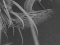
Spider foot
A picture of a mite spiders foot at a higher resolution than the gigapan that we took. This picture is at 3000x while the gigapan was 1000x.
Thursday, May 21, 2009
More on Zinc Oxide
David Speck wrote with more on Zinc Oxide
I thought ZnO crystals were a well known demo specimen for SEM work. At least when I was at Cornell in 1975, they were a favorite item for demonstrating the power of a SEM. I remember going to the SEM lab on Olin Hall with a friend who had bought some cheap SEM time at 2:00 AM. The most memorable item we looked at was the ZnO smoke sample.
Zinc burning in air forms perfect tetrahedral pointy crystals in a broad range of sizes that resemble a caltrop from outer space. See:
http://embedded.eecs.berkeley.edu/caltrop/pic/caltrop.jpg
I thought I'd have no trouble finding an image of these crystals, but an extended Google search didn't bring up a single example. Perhaps they have been forgotten. It would be interesting to see them again.
Tuesday, May 19, 2009
Moth Antenna
This morning we took a 135 image gigapan of the tip of a moth's antenna at 1,000x magnification.
The antenna curves in from the upper left.
View the full image at GigaPan.org
The antenna curves in from the upper left.
View the full image at GigaPan.org
Monday, May 18, 2009
Future Gigapan Ideas
This is in no way a comprehensive list, but what I have gathered so far is that people have expressed interest in seeing the following things imaged:
- Bee, leg/pollen
- Leaf, stoma
- Hard drive platter
- Soil
- Pond water
- Coca Cola
- Artificial sweeteners
- Bacon (cooked or uncooked?)
- Fingerprint
- Zinc oxide structures
- Cloaking material
- Blue Morpho butterfly wing
- Super lens
Possible subjects - Zinc Oxide crystals
David Speck wrote with a suggestion:
What do you think we should image?
You might want to try some zinc oxide crystals. They form amazing three dimensional tetrahedral structures. I saw them with a SEM a lifetime ago.Aside from the whole 'very toxic' thing this sounds fun.
You can make it by igniting a thin strip of zinc metal (rescued from the shell of a dead cheap carbon-zinc battery). It burns like magnesium, but with less enthusiasm.
The thick white smoke is very toxic to the lungs, so don't breathe it. It should stick to a glass microscope slide without much encouragement.
What do you think we should image?
Thursday, May 14, 2009
SEM Image of NaCl
An SEM gigapn of sodium chloride (NaCl), table salt. It was dissolved in water then left to condense back out on a piece of Silicon. This piece is about 1mm long and 0.5mm wide.
This is composed of 84 images taken at 1500X magnification.
View the full image at GigaPan.org
Taken by Jay Longson
You can pan and zoom in the image, or click on the blue title bar to jump to this gigapan on the gigapan.org site.
This is composed of 84 images taken at 1500X magnification.
View the full image at GigaPan.org
Taken by Jay Longson
You can pan and zoom in the image, or click on the blue title bar to jump to this gigapan on the gigapan.org site.
SEM Image of blood and hair
Seventy pictures at 1200x magnification of an eye lash (really an eyebrow hair) next to a dried blood sample using the SEM. You can see the red blood cells in the sample.
View the full image at GigaPan.org
Taken by Molly Gibson (who was also the blood and hair donor)
You can pan and zoom in the image, or click on the blue title bar to jump to this gigapan on the gigapan.org site.
View the full image at GigaPan.org
Taken by Molly Gibson (who was also the blood and hair donor)
You can pan and zoom in the image, or click on the blue title bar to jump to this gigapan on the gigapan.org site.
GigaPan Epic 10,000x
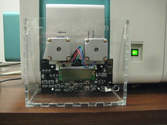
The nano Gigapan attached to the knobs on the Hitachi Scanning Electron Microscope.
The only glitch is that because there are no gears the stepper motors are turning the wrong way, so the picture numbering is off.
I think we just need to flip the polarity of the two pairs of wires to each stepper and it will magically move the right direction.
Click on the photo to enter Jay's photostream on flickr where you can see other pictures of the nano Gigapan mounted to the SEM.
Prototype for the nano gigapan
April 22, 2009 - This is the first pass Nano gigapan unit. Jay designed this to be cut out on the laser cutter at Tech Shop. We found some mat board for this first prototype. You can see all of the burnt edges. There was ash flying all over!
You can see more of these pictures in Rich's nanogigapan set on flickr
You can see more of these pictures in Rich's nanogigapan set on flickr
The Ant
This is the first proper nano-gigapan using the a modified gigapan unit attached to a scanning electron microscope (SEM). The image was then assembled was then stitched using the gigapan stitching software. The image is of an ants head at 1000X magnification. It took 64 images.
View the full image at GigaPan.org
View the full image at GigaPan.org
Wednesday, May 13, 2009
Welcome to the Nano Gigapan Pan
From the beginning the GigaPan project has been about reaching out, exploring, and connecting people with each other and with the wonderful shared world around us.
In that spirit Jay Longson has created the 'Nano Gigapan.' This is GigaPan modified to control a Scanning Electron Microscope.
Follow this blog to see our progress at exploring the world at the (near) nano scale!
In that spirit Jay Longson has created the 'Nano Gigapan.' This is GigaPan modified to control a Scanning Electron Microscope.
Follow this blog to see our progress at exploring the world at the (near) nano scale!
Subscribe to:
Comments (Atom)


